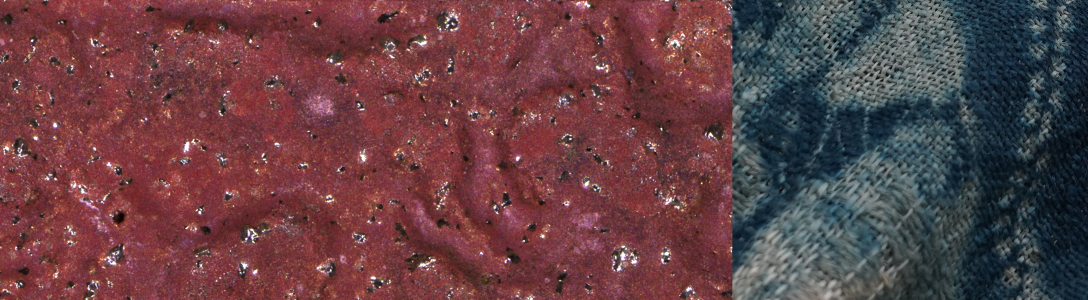

Here are some images from my first electron microscopy session with a sample from the July wood firing at Cobb Mountain Art & Ecology Center. These were all acquired using the Magellan Scanning Electron Microscope (SEM) in the Stanford Nano Shared Facilities, and the sample is the same one shown in color optical microscopy images from a previous post.
I prepared an SEM sample by cutting into the lid with a Dremel tool and diamond saw wheel; the cut-out piece mounted on a metal stub is shown here in an optical microscope montage:
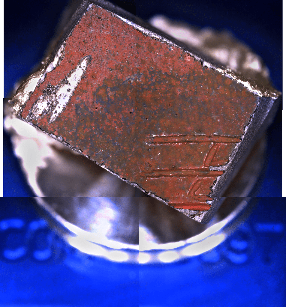
The image “stitching” here is unfortunately not soo good — the Zeiss Smartzoom5 has been slipping out of stage alignment but I’m hoping to recalibrate it soon once Zeiss sends me a part I need. This piece was taken from an area near the upper-right corner of the “ROI1” images in the Microscopy 1 blog post from 21 July. On the left side you can see a big scuff mark where I unfortunately slipped with the Dremel tool and ground off a bit of the nice red surface layer, and you can see some chipping of the surface layer around the edges of the piece (revealing the grey bulk clay underneath). As I have discussed elsewhere, there’s a very interesting and complex story about how the blacks and reds form as a thin surface crust on clays fired in wood-burning kilns with reduction cooling. As I’ll show in an upcoming post there are some images from this sample that provide new evidence in support of our current theory about how these reduction-cool colors form, but in this post I’ll just provide some survey images to introduce you to the micro- and nano-scale “scenery” of a wood fired ceramic surface as revealed by SEM.
The following three images show a patch of the sample surface at successively higher magnifications — 150x, 1500x and 12000x — with scale-bars in the lower right corner of each frame:
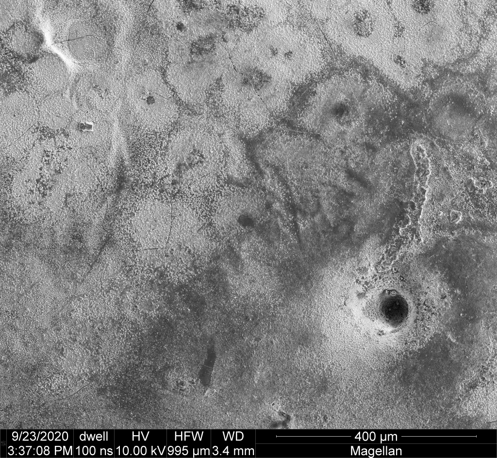


The topographic features visible in this type of SEM image generally correspond to micro/nano-crystals that form as the ceramic surface cools in the kiln after being fired to temperatures in the range of 2400F/1300C.
I’ve been seeing the best evidence for the detailed formation mechanisms of such crystals — and especially the specific iron oxide crystals responsible for the reduction cool red colors — in spots where the red iron crystals are relatively sparse, revealing the glassy black subsurface that they grow up from. The following two images show details of such a spot:
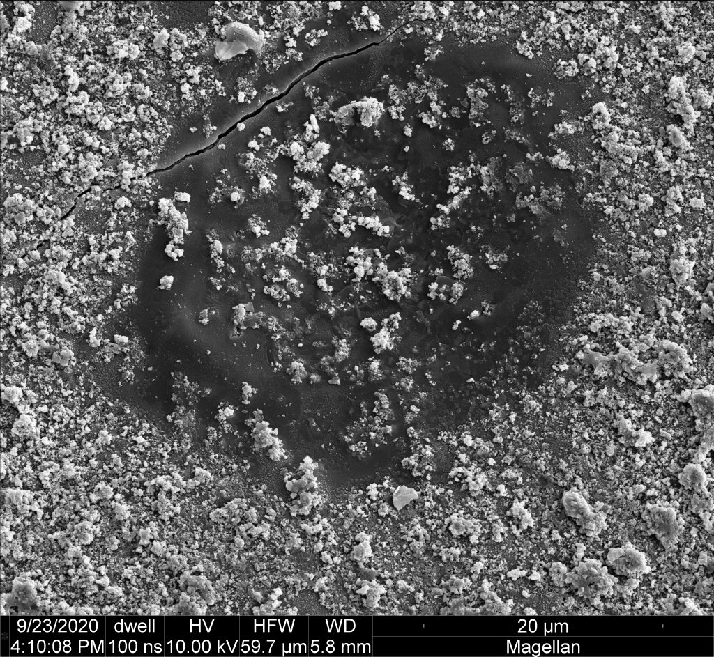

Here the right image is a closeup of the left.
The following three images show a region near the edge of the sample at successively higher magnifications, zooming in on what appears to be the inside surface of a bubble in the glassy ceramic surface that was broken open by chipping from the Dremel tool:


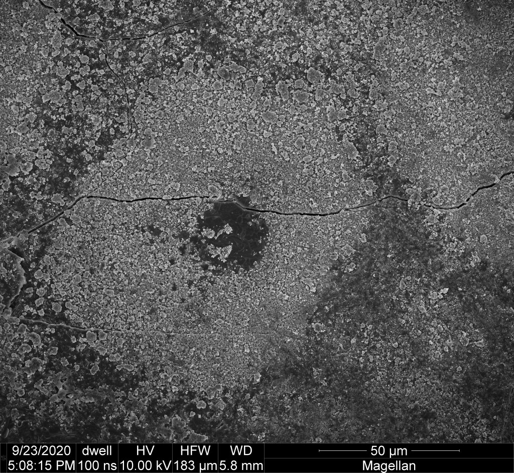
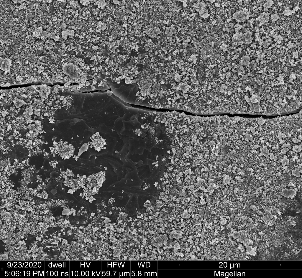
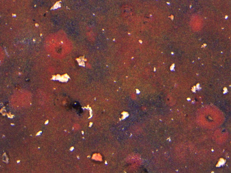
And finally (for now) an additional set of three images of a red halo around a black glassy spot, such as the ones visible in an optical microscope image from the 11 July blog post, which I’ll repost after the SEM images. The surface areas covered in “piles” of small angular crystals (hematite) are red to the eye, while the central glassy patch (an alkali-aluminosilicate glass of some kind) appears black.


[…] SEM study (see “microscopy 2”) with the sample cut from the wood-fired lid has provided some images that provide novel evidence […]
LikeLike