
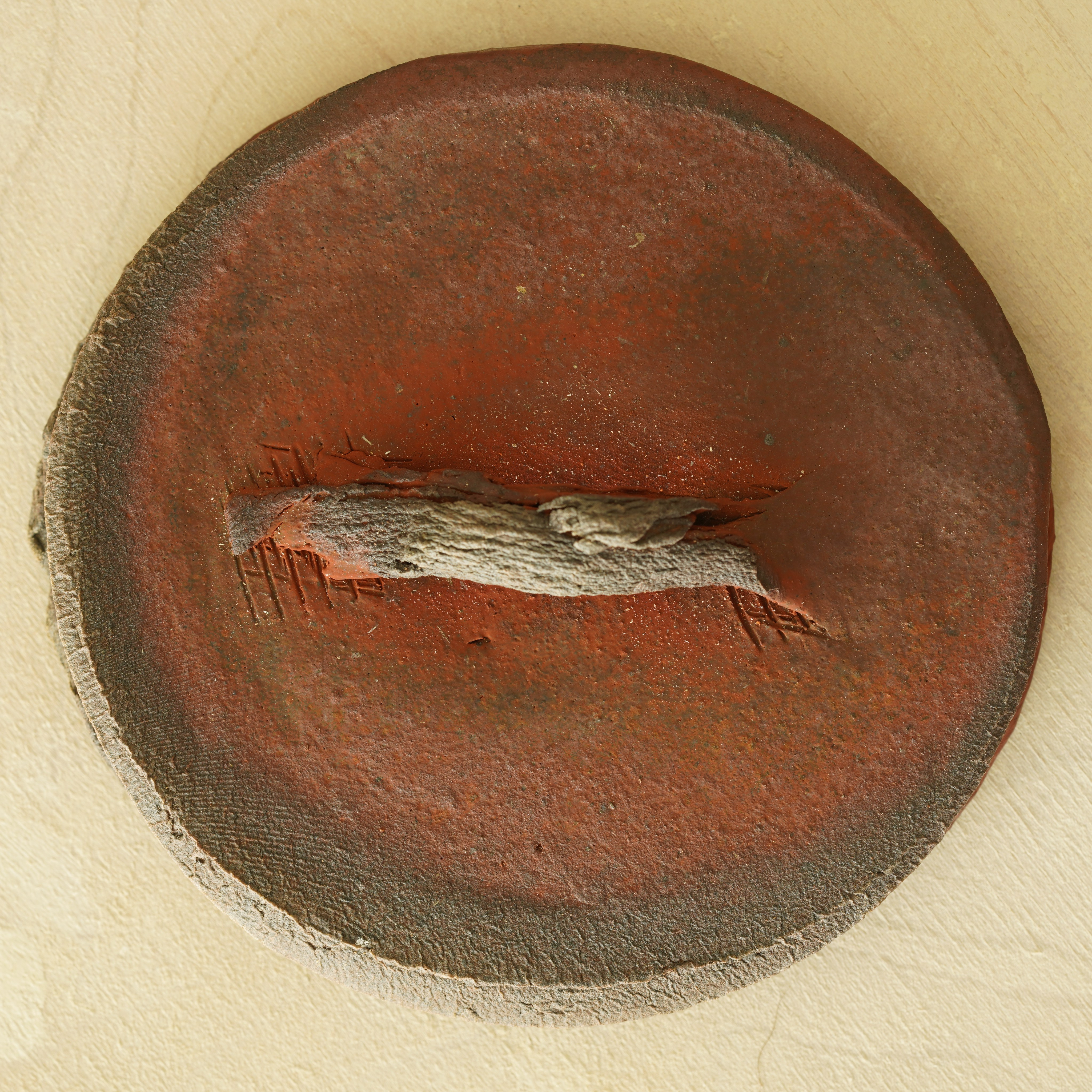
Yesterday I started doing some survey optical microscopy to help select a sample or two to prepare for electron microscopy. It tends to work best to start with a piece that’s already relatively flat, so I immediately thought about the top surface of the lid of this container:


The above photos were just taken with my cell phone; here’s a better photo of the lid top surface, taken using a digital camera with a high-quality 90mm macro lens:
Here’s a crop of the above digital camera photo, showing a region that I’ll call ROI1, near the bottom-left of the “handle”:

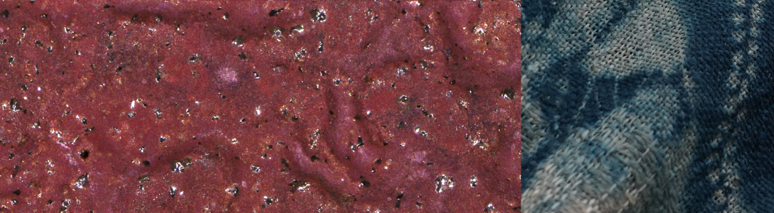
The above was taken in indirect daylight, against a light background, with the exposure set to the bright side. The next image shows a montage of EDF (extended depth-of-field) images acquired on our Ziess Smartzoom5 optical microscope, of roughly the same ROI1:

This microscope image is taken with direct LED ring-light illumination, against a dark background. It must be said the daylight digital camera image looks more true to color in terms of what one sees by eye, however, the microscope illumination setup is better for detailed studies in that it is much more precisely reproducible from one sample/image to the next. In both images it is clear that there are a range of purple-to-scarlet shades of what we would call patches of reduction-cool red (I’ve written about these on another website). The above microscope image was taken at a nominal magnification of 34x; here’s another montage of a smaller region within ROI1 taken at 155x (this is quite a large high-res file; to view in full resolution you’ll need to download the file and use an image viewer on your computer — click on the image here and then save the target file from the “view full size” button in the lower-right):

Now an even smaller sub-region of interest, on the left as seen at 34x by the Zeiss microscope and on the right as a digital camera photo (with slightly different orientation) taken with a 20mm Zongyu super-macro lens:


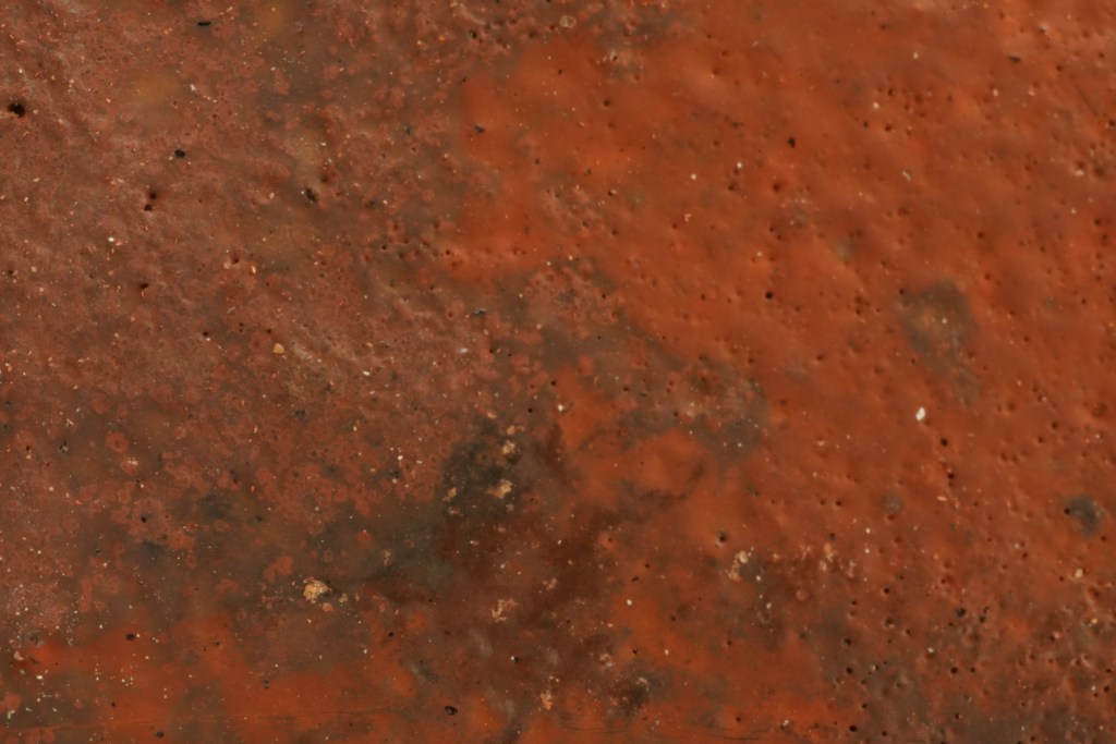
And now within this, a final zoom-in to 334x:

In this series of images I am interested in the way that red halos appear around what look like pinholes in the surface — this will make a nice focus for study with the electron mciroscope. Note that this last 334x image was acquired using a special glare-reduction feature of the Zeiss microscope, in which the angle of LED illumination is varied and an image synthesized in a way that minimizes specular reflections. The video below shows what the microscope looks like when operating in this mode:
Here’s a gallery of additional surface details from the lidded container, all taken with the digital camera and the 20mm Zongyu super-macro lens:


The 20mm super-macro is a nice lens to have, for capturing color tones in indirect natural light. It’s a bit tricky to use as the working distance is quite small:


[…] Here are some images from my first electron microscopy session with a sample from the July wood firing at Cobb Mountain Art & Ecology Center. These were all acquired using the Magellan Scanning Electron Microscope (SEM) in the Stanford Nano Shared Facilities, and the sample is the same one shown in color optical microscopy images from a previous post. […]
LikeLike